


As an Amazon Services LLC Associates Program participant, we earn advertising fees by linking to Amazon, at no extra cost to you.
Microscopic Insights into Moss Ecosystems
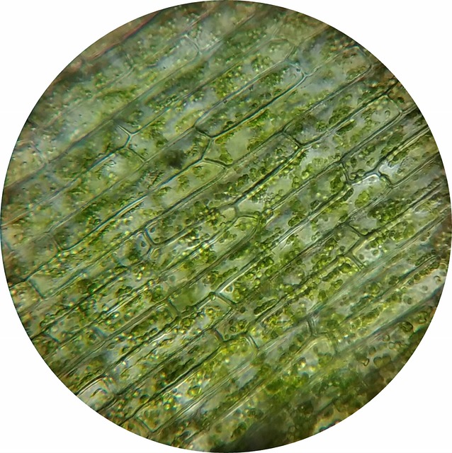
Many people overlook mosses, but they’re vital players in ecosystems. Microscopy reveals their hidden structures and functions. Observing moss under a microscope uncovers how they reproduce and thrive in various environments.
Most researchers stick to traditional microscopy. I believe we should embrace advanced techniques like confocal microscopy. This method enhances clarity, allowing us to see moss in three dimensions, revealing details that light microscopy misses.
According to B. C. Stagg and J. Dillon, “The intricacies of moss and their role in ecology can sometimes only be appreciated through the detailed observations enabled by microscopy.” This insight drives home the importance of microscopic studies.
Another innovative approach is using atomic force microscopy. It analyzes surface properties of mosses, giving us insights into their resilience. This is a game changer for understanding how mosses adapt to their environments.
As mosses face threats like habitat loss, integrating microscopy into conservation strategies is crucial. Understanding their biology at a cellular level can inform effective restoration practices. Microscopic assessments could be key to monitoring moss health and biodiversity.
Engaging in this kind of research isn’t just for scientists. Citizen science projects can involve communities in studying moss ecosystems. This not only broadens data collection but also nurtures a love for our local flora.
Let’s not underestimate the power of microscopy in revealing the complex roles of mosses. They’re more than just green ground cover; they’re essential to ecosystem health.
Apr 11, 2019 … Currently, researchers often use fluorescence microscopy to examine the structure and growth of plants, but this technique is limiting for …
Plant Disease Library: How To Methods, Use a Compound Microscope, Making a Moist Chamber, Making a Slide, Using Stain, Making a Wet Mount Slide, Submit a …
Dec 8, 2016 … “Stomatal guard cells are fascinating because they are some of the most dynamic cells in plants,” said Anderson. “Unlike most plant cells, which …
Grants from NSF power super-resolution microscope, probe cell …
Apr 16, 2024 … CAMBRIDGE, Mass., April 16, 2024—The famous Ware Collection of Blaschka Glass Models of Plants (the Glass Flowers) includes a series of models …
New in the Glass Flowers gallery: The Blaschkas at the Microscope …
Jul 16, 2014 … Technology best of its kind on campus. Giardia flagella. By Karyn Houston Plant & Microbial Biology. A microscopy expert at UC Berkeley has …
CNR Purchases Powerful New Microscope | Plant & Microbial Biology
Understanding Plant Microscopes and Their Importance
Microscopes are the unsung heroes of plant research. They unveil hidden details that can change our understanding of plant biology. For instance, the intricate structures of plant cells are only visible under a microscope. This insight is vital for fields like agriculture and ecology.
Many believe traditional light microscopes are the only option. I think alternatives like scanning electron microscopy (SEM) should be more widely used. SEM provides stunning three-dimensional images that reveal textures and surface structures of plants.
According to Sophie Cox from the Duke Research Blog, “Seeing tiny creatures under a microscope is a powerful, intoxicating thrill.” This thrill is not just for fun; it deepens our ecological understanding.
Moreover, fluorescence microscopy is a groundbreaking technique. It allows scientists to observe living plant cells in real-time, shedding light on cellular processes. This dynamic approach can transform our understanding of plant responses to environmental changes.
Some argue that digital microscopy is the future. However, I think we should not overlook the value of traditional methods. They offer a hands-on experience that is irreplaceable in education and research.
Incorporating artificial intelligence into microscopy analysis is another exciting frontier. It could lead to faster species identification and classification, making research more efficient.
In summary, embracing diverse microscopy techniques will significantly enrich our understanding of plant life. The future of botanical research is bright, thanks to these powerful tools.
The Role of Citizen Science in Microscopy
Citizen science is shaking things up in microscopy. Many think it’s just about data collection. But I believe it’s about community engagement and education.
Projects like Fjord Phyto invite everyone to gather samples. This isn’t just busywork; it’s real science! Volunteers analyze phytoplankton under microscopes, contributing to vital research. According to Daniel Brooks from Swoop Antarctica, “Engaging with citizen science adds significant value to our understanding of ecosystems.”
Most folks assume that only trained scientists can handle microscopy. I think that’s a narrow view. With the right tools, anyone can explore the microscopic world. Imagine a mobile microscopy lab visiting schools! This hands-on approach could spark curiosity and deepen understanding.
Another angle? Integrating technology with citizen science. Apps could guide volunteers through sample collection and analysis. This way, they’re not just participants; they’re contributors to real-time data collection.
And let’s not overlook the educational aspect. Citizen science can lead to workshops that teach microscopy skills. This builds a knowledgeable community that values science. As we see more citizen science initiatives, we’re not just gathering data; we’re cultivating a culture of inquiry.
Let’s embrace this shift. Citizen science isn’t just a trend; it’s a powerful tool for advancing botanical research.
Microscopy Techniques for Plant Studies
Explore various microscopy techniques that enhance our understanding of plant biology and ecology. These methods reveal hidden details and foster innovative research.
- Light microscopy is the go-to for many plant researchers. It provides a basic view of cellular structures and functions.
- Scanning electron microscopy (SEM) offers a stunning, three-dimensional perspective. It uncovers surface details that light microscopy misses.
- Confocal microscopy allows for precise imaging of plant tissues. This technique enhances clarity by scanning layers of specimens.
- Atomic force microscopy analyzes surface properties. It reveals how mosses adapt to their environments.
- Fluorescence microscopy studies living plant cells in real-time. It highlights specific structures, offering dynamic insights into cellular processes.
- Digital microscopy integrates technology with traditional methods. It simplifies image capture and analysis, making research more accessible.
- Citizen science projects expand the reach of microscopy. They engage communities in botanical research, collecting valuable data.
- Mobile microscopy labs bring research to local communities. They offer hands-on training and immediate application of microscopy techniques.
- Microscopic studies of moss can inform conservation strategies. Understanding their biology at a cellular level is key to protecting these species.
- The integration of artificial intelligence in image analysis is on the rise. It promises breakthroughs in identifying and classifying plant species.
Future Directions in Plant Microscopy Research
Most experts think fluorescence microscopy is the pinnacle of plant research. I believe we should push boundaries further by integrating artificial intelligence for image analysis. This combination can lead to breakthroughs in species identification that traditional methods miss.
Many believe that studying plant cells is limited to static images. But I argue that real-time imaging of living cells offers a dynamic view of plant physiology. It’s that simple—seeing processes as they happen can change everything.
While some focus solely on cellular structure, I think we should also explore ecological interactions. Understanding how plants respond to their environments at a microscopic level can inform conservation efforts. This perspective shifts the focus from just observation to actionable insights.
As noted by Daniel Brooks from Swoop Antarctica, “Engaging with citizen science adds significant value to our understanding of ecosystems.” Imagine harnessing this community-driven approach to collect microscopic data. It could enhance research and foster local stewardship.
Let’s not forget about mosses. They often get overlooked, but microscopic studies can reveal their ecological importance. Understanding their role in water retention and soil formation is critical, especially in changing climates.
We should also explore the potential of mobile microscopy labs. Bringing these labs to communities can democratize plant research. It allows hands-on experiences that traditional labs can’t offer.
In summary, the future of plant microscopy lies in innovation and community engagement. By embracing new technologies and methodologies, we can unlock a deeper understanding of plant life.
Innovative Techniques for Plant Microscopy
Most people think traditional light microscopes are the only way to study plants. I believe that exploring techniques like scanning electron microscopy (SEM) can unlock a new level of understanding. SEM provides stunning three-dimensional images, revealing details that light microscopy might miss.
There’s also a growing interest in digital microscopy. This method captures images directly to a computer, making analysis easier and more efficient. Imagine being able to manipulate and analyze data on-the-fly!
Then there’s fluorescence microscopy. It allows scientists to observe living plant cells in real-time, illuminating specific structures or proteins. This dynamic insight is groundbreaking for understanding plant physiology.
While many researchers focus on traditional methods, I think incorporating artificial intelligence into image analysis could be revolutionary. AI can help classify plant species more accurately and quickly.
As technology continues to evolve, so should our approaches to plant microscopy. Embracing these innovative techniques can lead to remarkable discoveries in plant biology. Let’s not settle for the old ways when the future holds so much potential!
According to Daniel Brooks from Swoop Antarctica, “Engaging with citizen science projects empowers individuals to make significant contributions to our understanding of complex ecosystems.” This reinforces the idea that we should keep pushing the boundaries.
Jun 20, 2024 … … Microscope may be a vital tool in the advancement of your research. To learn more about CytoViva's Enhanced Darkfield Hyperspectral Microscopy …
Sep 1, 2021 … This is confused with the industrial term wood fibers, which means a mash-up of all the wood cells to make wood pulp. In plant science, the …
Key Applications of Plant Microscopy
Plant microscopy opens up a world of insights into plant biology and ecology. Here are some key applications that showcase its importance.
- Microscopy reveals hidden structures. It allows us to see plant cells and tissues in detail, enhancing our understanding of plant biology.
- Disease identification is crucial. By examining plant samples under a microscope, researchers can spot diseases early, which is vital for agriculture.
- Moss studies benefit greatly. Microscopes help us observe the intricate structures of moss, shedding light on their ecological roles.
- Fluorescence microscopy is a game changer. This technique allows scientists to visualize live plant cells and their processes in real time.
- Citizen science expands research. Community involvement in microscopy projects, like Fjord Phyto, broadens data collection and engages the public.
- Alternative microscopy techniques enhance insights. Methods like atomic force microscopy provide unique views of plant surfaces, revealing mechanical properties.
- AI is revolutionizing image analysis. Integrating artificial intelligence into microscopy can streamline data processing and improve species identification.
- Educational programs foster awareness. Hands-on microscopy workshops can ignite interest in plant science among students and the community.
single celled organisms, bugs, viruses, plants, cells, etc. ⬤. ⬤. beautiful colorized images. Microangela's Electron Microscope Image gallery, single celled …
Identification of components and toxic substances, such as plants and plant parts, algae, insects, or bait material. … Site Information. Website Feedback …
Toxicology Evaluation (Microscopy) – Texas A&M Veterinary Medical …
Oak Wilt spores under the microscope. For more information about Oak Wilt, see Oak Wilt in Minnesota or the DNR page for Oak Wilt.
… plant & animal microtechnique, histology, and cryotomy. IOM112024 malaria … The Golub Microscope Collection: Web site dedicated to Dr. Orville J …
The Biological Imaging Facility – Core microscope facility at UC …
Scanning Electron Microscopy · High resolution FE-SEM · Immuno-SEM · Array tomography SEM, CLEM …
What types of microscopes are best for studying plants?
Many people think light microscopes are sufficient for plant studies. I believe using advanced techniques like scanning electron microscopy (SEM) is far superior. SEM provides high-resolution images and 3D views of plant structures, revealing details that light microscopes miss.
Some might argue that traditional methods are enough. However, I find that integrating fluorescence microscopy allows us to observe living cells in real time. This dynamic insight can significantly deepen our understanding of plant physiology.
Moreover, combining microscopy with artificial intelligence for image analysis could revolutionize how we classify and study plant species. This innovative approach can lead to breakthroughs that traditional methods simply can’t achieve.
As noted by Daniel Brooks from Swoop Antarctica, “Engaging with citizen science adds significant value to our understanding of ecosystems.” This perspective highlights the need for diverse methodologies in plant microscopy.
What insights can microscopy provide about moss species?
Microscopy reveals the intricate structures of mosses that are often missed by the naked eye. Most people think traditional light microscopy is sufficient for studying moss. But I believe using confocal microscopy offers a clearer, three-dimensional view of these fascinating organisms.
Through microscopy, we can observe how moss captures water and nutrients. This is vital for understanding their role in ecosystems. According to B. C. Stagg and J. Dillon, “The intricacies of moss and their role in ecology can sometimes only be appreciated through the detailed observations enabled by microscopy.”
Moreover, atomic force microscopy can uncover the mechanical properties of moss. This helps us learn how they adapt to various environments. These insights are crucial for conservation efforts as mosses face habitat loss.
How can citizen science contribute to microscopy research?
Most people think citizen science is just about collecting samples. But I believe it’s way more impactful. It empowers communities to engage directly with scientific research.
Take projects like Fjord Phyto. Volunteers gather phytoplankton samples from Antarctic waters. These samples are then analyzed under microscopes, leading to significant discoveries. According to Daniel Brooks from Swoop Antarctica, “Engaging with citizen science adds significant value to our understanding of ecosystems.”
Integrating mobile microscopy labs could take this a step further. Instead of just collecting data, these labs can offer hands-on training to local communities. This approach fosters immediate application of microscopy techniques, creating lasting skills.
Let’s not forget the potential of combining citizen science with educational programs. Imagine students learning about plant biology while contributing to real research! It’s a win-win for both education and science.
Most folks think traditional light microscopes are enough. I believe we need to embrace advanced techniques like fluorescence microscopy. This method lets us observe living plant cells in real time, revealing dynamic cellular processes.
Many researchers focus on conventional methods, but I argue that integrating artificial intelligence in image analysis can lead to breakthroughs. It’s not just about seeing; it’s about understanding plant responses to environmental changes.
As Sophie Cox said, “Seeing tiny creatures under a microscope is a powerful, intoxicating thrill.” It’s time we amplify that thrill with cutting-edge technology!
For more insights, check out the work of Daniel Brooks on citizen science.
Microscopes uncover the unseen layers of plant life. They show us how cells function and interact. It’s fascinating to witness the intricate details of plant cells.
Most people think traditional light microscopes are the only way to go. But I believe that techniques like fluorescence microscopy bring a whole new perspective. They let us observe living cells in real time, revealing dynamic processes.
According to Daniel Brooks from Swoop Antarctica, “Engaging with citizen science adds significant value to our understanding of ecosystems.” This highlights how community involvement can enhance our insights.
Moss studies are particularly enlightening. They show how these plants capture water and nutrients, crucial for their survival. Using advanced microscopy can shed light on their ecological roles.
New methodologies in microscopy could redefine our approach to plant biology. The integration of artificial intelligence in analyzing microscopic data is one exciting frontier.
Most people think citizen science just gathers data. I believe it transforms research by engaging communities directly. Projects like Fjord Phyto invite volunteers to collect phytoplankton samples. This hands-on approach opens doors to discoveries that traditional methods might miss.
Engagement boosts awareness about ecosystems. As Daniel Brooks from Swoop Antarctica said, “Engaging with citizen science adds significant value to our understanding of ecosystems.” It’s that simple!
We can also explore mobile microscopy labs. They bring the lab to the community, allowing for training and immediate application. This direct mentorship enhances accuracy and involvement.
As an Amazon Services LLC Associates Program participant, we earn advertising fees by linking to Amazon, at no extra cost to you.

I’ve always been captivated by the wonders of science, particularly the intricate workings of the human mind. With a degree in psychology under my belt, I’ve delved deep into the realms of cognition, behavior, and everything in between. Pouring over academic papers and research studies has become somewhat of a passion of mine – there’s just something exhilarating about uncovering new insights and perspectives.