As an Amazon Services LLC Associates Program participant, we earn advertising fees by linking to Amazon, at no extra cost to you.
Integration of Digital Imaging in Veterinary Practices
Exploring the innovative shift towards digital imaging in veterinary microscopy, highlighting its benefits and new approaches.
- Digital imaging is reshaping veterinary diagnostics. It offers clearer images for better analysis.
- Many believe traditional methods are sufficient. I think digital tools provide insights that can’t be matched.
- According to UC Davis, “Researchers developed a new microscope to capture high-speed images of brain cell activity with less harm to brain tissue.” This shows the potential for less invasive techniques.
- Real-time imaging can transform how we diagnose. It allows for immediate insights into animal health.
- Most practitioners rely on established techniques. I argue that embracing digital microscopy can lead to earlier disease detection.
- Incorporating AI into imaging processes could enhance accuracy. This advancement could streamline workflows and improve patient outcomes.
- Some think digital imaging is just a trend. I believe it’s the future of veterinary medicine, with endless possibilities.
- Collaboration among professionals can be improved. Digital tools facilitate sharing insights and findings across platforms.
Types of Veterinary Microscopes for Different Applications
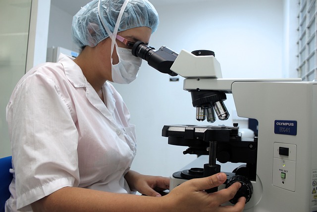
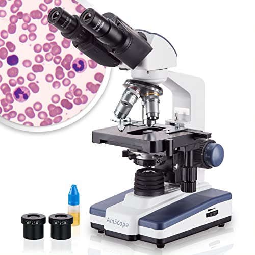
Veterinary microscopes come in various types, each tailored for specific diagnostic needs. The compound microscope is a staple, magnifying up to 1000x. It’s perfect for blood analysis and cytology.
Then we have stereo microscopes. They provide 3D views, making them great for larger specimens. These are ideal for dissections and examining tissue samples.
Digital microscopes are changing the game. They connect to computers for enhanced analysis and easy data sharing. This feature is super helpful for remote consultations.
Most people think traditional optical microscopes are enough. But I believe portable field microscopes are the future. They allow immediate diagnosis in outdoor settings, which is a game changer for wildlife health assessments.
According to Hery Ríos-Guzmán from Cornell University, “Veterinarians have a wide array of tools that can be used to enhance the incredible work that biologists and other conservationists do.” This highlights the importance of having the right tools for every situation.
As technology advances, the integration of digital imaging is essential. It enhances image quality and accessibility, making veterinary diagnostics more efficient. This shift is not just a trend; it’s a necessity for modern veterinary practices.
The Georgia Electron Microscopy Facility provides diagnostic services to the veterinary and medical professions. We have the unique opportunity to work …
… Microscope at the College of Veterinary Medicine. View the event schedule below. Register to attend. Welcome and Introduction Becky Williams, PhD – Director …
Introducing a New Confocal Microscope at the College of Veterinary …
This upright widefield microscope is equipped with both color and monochrome cameras, making it suitable for imaging either histology slides or fluorescently …
Ingrid Brust-Mascher uses a Leica Super High-Resolution Confocal Microscope in the Health … Future Veterinary Medical Center · Campus Directory. #1 Vet …
Microscopy Core Facility · Instruments. The Multi Camera Array Microscope (MCAM) Kestrel can provide image capture and analysis of high throughput chemical and …
Challenges and Future Directions in Veterinary Microscopy
Veterinary microscopy is at a crossroads. Many believe that traditional methods are sufficient for diagnostics. But I think we need to embrace new technologies to improve outcomes.
While conventional microscopes have served us well, integrating AI and automation could enhance diagnostic accuracy. Imagine machines analyzing slides faster than any human could!
Investing in high-throughput imaging systems seems daunting. But the benefits of precision and speed are undeniable. As noted by experts, “The increased precision and speed offered by modern systems promise a significant improvement in diagnostic capabilities.”Source.
It’s not just about tools; it’s about knowledge. Continuous professional development is crucial. We must adapt to new technologies to stay relevant.
While most practitioners focus on traditional approaches, I believe interdisciplinary methods could transform our field. Incorporating data science might lead to groundbreaking algorithms for image analysis.
Ethical considerations are also paramount. We need to address how we handle specimens and the implications of advanced imaging technologies. This isn’t just about progress; it’s about responsible practices in veterinary medicine.
As we look to the future, let’s not shy away from these challenges. Instead, let’s tackle them head-on, ensuring that veterinary microscopy evolves to meet the needs of animal health.
Key Microscopy Techniques Used in Veterinary Diagnostics
Here’s a quick look at essential microscopy techniques that are game changers in veterinary diagnostics. Each technique offers unique insights that help veterinarians make informed decisions.
- . Bright Field Microscopy: This is the go-to for general histological evaluations. It uses transmitted light to visualize specimens, making it simple yet effective.
- . Fluorescence Microscopy: This technique allows us to view specific cell structures. Using fluorescent dyes, it highlights important cellular components.
- . Electron Microscopy: This one provides ultra-high resolution. It’s crucial for spotting tiny abnormalities, like viral infections.
- . Confocal Microscopy: It enhances imaging depth and detail. Perfect for real-time analysis of living tissue samples.
- . In Vivo Microscopy: This innovative method images living tissues without excision. It offers real-time insights into health dynamics.
- . Digital Imaging: This modern approach enhances image quality and accessibility. It allows for better data sharing among veterinary professionals.
- . Cryo-Electron Microscopy: This advanced technique captures samples at cryogenic temperatures. It’s essential for studying delicate structures without damage.
Importance of Veterinary Microscopes in Diagnosing Animal Health
Veterinary microscopes are indispensable tools for diagnosing animal health. They allow veterinarians to see what’s happening at a cellular level. This insight is crucial for identifying diseases and infections.
While traditional optical microscopes are the norm, I believe we should embrace emerging technologies. Digital pathology and AI are game changers. They can analyze large volumes of samples quickly, offering more detailed insights.
Most experts think that sticking with conventional methods is sufficient. But I argue that integrating these new technologies can enhance diagnostic accuracy significantly. Imagine being able to diagnose conditions faster, leading to better outcomes for our furry friends!
We’re at a turning point in veterinary medicine. The future of veterinary microscopy lies in innovation. By adopting new technologies, we can improve animal welfare and advance research.
As noted by Michelle Greenfield from Cornell University, “The ability to observe the microscopic structures helps in accurate diagnosis and facilitates timely medical intervention.” This is the essence of why we need to push boundaries.
Advanced Microscopy Techniques in Veterinary Medicine
Most people think traditional bright field microscopy is the gold standard in veterinary diagnostics. I think we’re missing out on the real game-changers—like fluorescence microscopy. This technique uses specific dyes to highlight cellular structures, making it easier to spot abnormalities.
Another exciting approach is electron microscopy. It provides ultra-high-resolution images, allowing us to identify minute details in cellular structures. According to Nancy D. Lamontagne from UC Davis, “By providing a tool that can observe neuronal activity in real time, our technology could be used to study the pathology of diseases at the earliest stages.” That’s a big deal!
Then there’s confocal microscopy, which adds depth to our imaging capabilities. It’s like looking at a 3D model rather than a flat picture. This technology is perfect for examining living tissues without the need for excision, giving us real-time insights.
Some experts believe that in vivo microscopy techniques are the future. They allow for dynamic imaging without invasive procedures. This could change how we diagnose and treat conditions in animals.
While many stick to conventional methods, I argue that embracing these advanced techniques can elevate our understanding of animal health. It’s not just about what we see; it’s about how we interpret it. The future of veterinary medicine lies in our ability to adapt and innovate.
To explore more about these techniques, check out the insights from UC Davis.
I generated clinical interests in the microscope by defining the necessity of this technology throughout veterinary clinics in the southeast. I… Show more. I …
Ashley Rogers – Office Manager – Lawrenceville-Suwanee Animal …
Jul 27, 2024 …Veterinary Microscopes Market 2024: 7.58% CAGR Growth Starting at USD 92 Billion in 2023, the "Veterinary Microscopes Market" is expected to …
Global Veterinary Microscopes Market Scope By Application | Trends
Common Applications of Different Types of Veterinary Microscopes
Veterinary microscopes are indispensable tools in animal health diagnostics. They serve various applications, each tailored to specific needs in veterinary practice. Here’s a quick rundown of how different types of microscopes are used in the field.
- Compound microscopes are the go-to for examining blood samples. They can magnify up to 1000x, revealing infections and abnormalities.
- Stereo microscopes provide a 3D view, making them perfect for larger specimens. They’re great for dissections and studying tissue samples.
- Digital microscopes integrate imaging technology for enhanced analysis. They allow for easy sharing and archiving of images, which is super helpful in consultations.
- Fluorescence microscopy uses specific dyes to highlight cellular structures. This technique is invaluable for identifying pathogens in tissues.
- Electron microscopy offers ultra-high resolution. It’s essential for spotting cellular abnormalities related to diseases like cancer.
- In vivo microscopy allows for real-time imaging of living tissues. This could change how we diagnose conditions without invasive procedures.
- Portable field microscopes are gaining traction. They enable immediate diagnostics in wildlife and outdoor settings, speeding up veterinary responses.
Use the Find a Test page to browse and search for available tests. Toxicology Evaluation (Microscopy) …
Toxicology Evaluation (Microscopy) – Texas A&M Veterinary Medical …
… microscope. The facility offers consultation, training, and support … This site is officially grown in SiteFarm.
Need a different test? Use the Find a Test page to browse and search for available tests.
Electron Microscopy – Texas A&M Veterinary Medical Diagnostic …
May 18, 2022 … Transmission electron microscopy of thin-sectioned tissues; Collaboration with researchers on creative scientific investigations; On-site …
California Animal Health & Food Safety Lab System – Electron …
vet looking through a microscope. Lab Skills. Students learn microbiological … This site is officially grown in SiteFarm.
Ethical Considerations in Veterinary Microscopy
Ethics in veterinary microscopy? Oh, absolutely! We’re talking about handling animal specimens with care and respect. Every sample tells a story, and we must honor that.
Most folks think that as long as the results are accurate, the methods don’t matter. But I think that’s a narrow view. We need to consider the implications of advanced imaging technologies on animal welfare.
For instance, the use of in vivo microscopy offers a non-invasive way to observe living tissues. This could change the game in how we approach diagnostics. We should prioritize techniques that minimize harm to animals.
Ethical considerations must also extend to data sharing. With digital imaging, how do we protect sensitive information? Transparency is key, but so is confidentiality.
Moreover, some argue that the push for innovation can overshadow ethical practices. But I firmly believe that ethics should guide our advancements. Innovation without ethics is a recipe for disaster.
As we explore these new frontiers, we must keep our moral compass intact. Let’s ensure that our pursuit of knowledge doesn’t compromise our values.
In the end, the conversation around ethics in veterinary microscopy is just beginning. We have a responsibility to lead with integrity.
Emerging Technologies in Veterinary Microscopy
Explore how new technologies are shaping the future of veterinary microscopy.
- Digital pathology is transforming diagnostics. It allows for faster analysis and improved accuracy in identifying diseases.
- AI integration can enhance diagnostic efficiency. Algorithms can analyze large sample volumes quickly, offering insights that may be missed by human eyes.
- In vivo microscopy is a game changer. It enables real-time imaging of living tissues, revolutionizing how we understand animal health.
- Portable field microscopes are gaining traction. They allow veterinarians to conduct immediate assessments in wildlife or outdoor settings.
- High-throughput imaging systems are on the rise. These systems promise to increase diagnostic precision and speed, but require investment and training.
- Automation in microscopy is becoming essential. It streamlines workflows, allowing veterinarians to focus on patient care rather than repetitive tasks.
- Ethical considerations are paramount. As technology advances, we must ensure responsible practices in handling animal specimens.
What are the main types of veterinary microscopes?
Veterinary microscopes come in various types, each with unique applications. The compound microscope is the most common. It offers high magnification, making it perfect for blood analysis and cytology.
The stereo microscope provides a 3D view, ideal for larger specimens. It’s great for dissections, giving a broader perspective.
Digital microscopes take it further, allowing image capture and analysis on computers. This is a game changer for remote consultations and educational purposes.
Some experts advocate for portable field microscopes, which enable immediate diagnosis in the field. This is essential for wildlife health assessments and rapid response to emerging issues.
As Hery Ríos-Guzmán from Cornell University states, “Veterinarians have a wide array of tools that can be used to enhance the incredible work that biologists and other conservationists do.” See more here.
How do advanced microscopy techniques benefit animal health?
Most people think advanced microscopy techniques are just fancy tools. I believe they’re game-changers for animal health diagnostics. These techniques, like fluorescence microscopy, allow us to see specific cellular structures, enhancing our understanding of diseases.
Traditional methods can miss critical details. With electron microscopy, we can identify ultrastructural changes in tissues. This precision is vital for diagnosing complex conditions.
Some argue that in vivo microscopy is the future. It offers real-time insights into living tissues, which could transform how we approach veterinary medicine. By observing dynamic processes, we can catch issues before they escalate.
Incorporating these advanced techniques into veterinary practices is essential. They not only improve diagnostic accuracy but also support better treatment plans. As Nancy D. Lamontagne from UC Davis said, “By providing a tool that can observe neuronal activity in real time, our technology could be used to study the pathology of diseases at the earliest stages.”
What challenges does veterinary microscopy currently face?
Many believe that the biggest challenge in veterinary microscopy is keeping up with new technologies. I think the real issue is the lack of training for veterinarians in these advanced tools. Without proper education, even the best equipment can go underutilized.
Financial constraints are another major hurdle. Most clinics can’t afford the latest high-throughput imaging systems. This limits their diagnostic capabilities and ultimately affects animal health.
Moreover, there’s a pressing need for interdisciplinary collaboration. Most people think traditional methods are sufficient, but I argue that integrating data science could lead to breakthroughs in diagnostic efficiency.
As highlighted by experts, embracing automation in microscopy could streamline workflows. Yet, many practitioners resist change, fearing it might replace their roles. This mindset needs to shift for the benefit of both veterinarians and their patients.
Incorporating ethical practices in handling specimens is essential. It’s not just about technology; it’s about responsible use of these advancements in veterinary medicine.
Veterinary microscopes are vital for pinpointing health issues in animals. They allow us to see details that are otherwise invisible. For example, analyzing blood smears can quickly identify infections.
Many professionals think traditional microscopes are enough, but I believe integrating digital imaging is the future. This tech not only improves image quality but also makes sharing findings easier.
According to UC Davis, new microscope technologies can enhance our understanding of disease pathology. Imagine the possibilities!
Incorporating AI into microscopy can streamline diagnostics. This means faster results and better animal care.
Many folks think traditional microscopy is enough for diagnosing animal diseases. But I believe advanced techniques like fluorescence and electron microscopy open new doors. They reveal details we often miss with standard methods.
For example, fluorescence microscopy highlights specific cell structures, making it easier to identify infections. According to Nancy D. Lamontagne of UC Davis, “By providing a tool that can observe neuronal activity in real time, our technology could be used to study the pathology of diseases at the earliest stages.”
Additionally, in vivo microscopy is being researched. Imagine real-time insights into living tissues without cutting them open! This could change how we approach diagnostics.
Veterinary microscopes are not one-size-fits-all. Each type serves unique diagnostic needs. For instance, compound microscopes excel in cellular analysis.
Many believe that traditional optical microscopes are sufficient. I argue that digital microscopes open new doors for remote diagnostics and collaboration.
Stereo microscopes offer depth perception for larger specimens. They are essential for dissections and detailed examinations.
With advancements, portable field microscopes are gaining traction. They allow immediate diagnosis in the field, which is a game changer for wildlife health assessments.
As highlighted by Hery Ríos-Guzmán from Cornell University, “Veterinarians have a wide array of tools that can be used to enhance the incredible work that biologists and other conservationists do.” Check out more from Cornell University.
Most people think traditional training suffices for veterinarians. I believe continuous learning is essential because technologies evolve rapidly. Veterinary professionals must adapt to stay relevant.
Emerging tools like AI and digital imaging transform diagnostics. Relying solely on past knowledge limits potential. According to Nancy D. Lamontagne from UC Davis, “By providing a tool that can observe neuronal activity in real time, our technology could be used to study the pathology of diseases at the earliest stages” source.
Portable field microscopes are gaining traction, enabling immediate diagnosis. This shift demands new skills and knowledge. We need to embrace these advancements to improve animal health.
As an Amazon Services LLC Associates Program participant, we earn advertising fees by linking to Amazon, at no extra cost to you.

I’ve always been captivated by the wonders of science, particularly the intricate workings of the human mind. With a degree in psychology under my belt, I’ve delved deep into the realms of cognition, behavior, and everything in between. Pouring over academic papers and research studies has become somewhat of a passion of mine – there’s just something exhilarating about uncovering new insights and perspectives.


